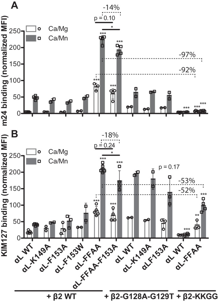
Figure 4.

Effect of the αI α1-helix mutations on αLβ2 conformational change. HEK293FT cells were transfected with the indicated combination of αL and β2 constructs. The cells were incubated with mAb m24 (A) that reports αLβ2 headpiece opening or KIM127 (B) that reports αLβ2 extension in the presence of 1 mm Ca2+/Mg2+ (Ca/Mg) or 0.2 mm Ca2+ plus 2 mm Mn2+ (Ca/Mn). The mAb binding was measured by flow cytometry and presented as the MFI normalized to αLβ2 expression measured by TS2/4 binding. The data are means ± S.D. (n ≥ 3; except for αl-K149A and αl-F153W, n = 2). Two-tailed t tests were used to compare the wild type and the mutants in the same conditions or as indicated. *, p < 0.05; **, p < 0.01; ***, p < 0.001. The numbers of percentages indicate the decreased levels of mAb binding.
