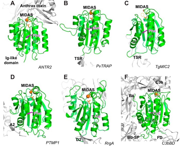
Figure 8.
Representative VWA structures. A–F, the crystal structures of VWA domains (in green) of human anthrax toxin receptor 2 bound with anthrax toxin (PDB code 1T6B) (A); P. vivax thrombospondin repeat anonymous protein (PDB code 4HQL) (B); T. gondii micronemal protein 2 (PDB code 4OKR) (C); blue mussel proximal thread matrix protein 1 (PDB code 4CN9) (D); S. pneumoniae pilus-related adhesion, RrgA (PDB code 2WW8) (E); and human complement C3b bound with factor B and factor D (PDB code 2XWB) (F). The DXSXS motif and metal ions of MIDAS are shown in orange. The residues of α1-helix that are equivalent to the Phe of integrin αI α1-helix are shown in magenta.

