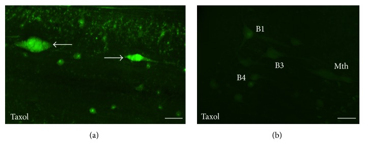
Figure 7.
Photomicrographs of dorsal views of the whole-mounted spinal cord (a) and brain (b) of a larval sea lamprey treated with Taxol showing the presence of FAM-LETD-FMK labeling in the tip of descending axons 1 week after treatment (arrows in (a)) and the absence of labeling in identifiable descending neurons of the brain (b). Rostral is to the right in (a) and to the top in (b). The ventricle is to the left in (b). B1, B3, and B4: Müller cells of the bulbar region; Mth: Mauthner cell. Scale bars: 40 μm in (a) and 50 μm in (b).

