
Figure 1.
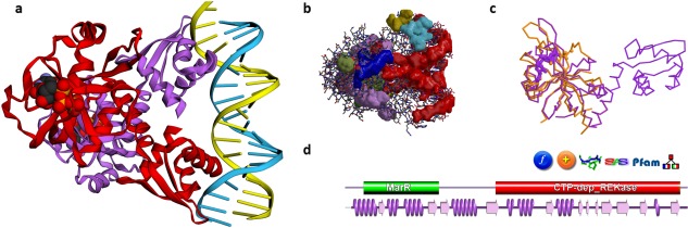
Some PDBsum analyses for PDB entry 5trd—a riboflavin kinase from Thermoplasma acidophilum. (A) Thumbnail image of the structure, as shown using 3Dmol.js on the entry's PDBsum page; (B) clefts in the structure, shown here as solid surfaces colored according to volume, with the largest shown in red; (C) 3D superposition based on a SAS sequence alignment between the 5trd protein, in purple, and, in orange, 2vbs, a partial structure comprising the first domain of riboflavin kinase from Methanocaldococcus jannaschii; d. domain representation of the 5trd protein, with the small purple wiring diagram underneath representing the protein's structural coverage, and the various icons above it leading to further analyses, as described in the main text.