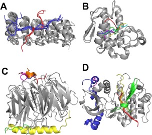
Figure 3.

Examples of different binding modes within one receptor cluster. (A) Cluster 28 (tankyrase and ankyrin), (B) Cluster 53 (phospholipase A2), (C) Cluster 11 (WD‐repeat), and (D) Cluster 13 (protein kinase). Receptors (gray) are bound to different peptides in different binding modes. Peptides within the same binding mode are shown using the same color. To simplify representation, only one peptide per binding mode and one receptor chain is shown in C and D. Representative PDB IDs for each receptor are, respectively: 3TWR,39 3JTI, 4J81,40 and 1NVS.41
