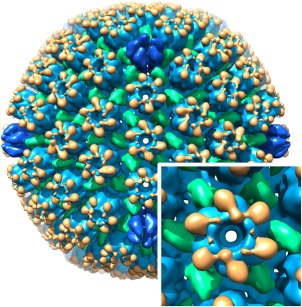
Figure 6.

Segmentation of the herpes simplex virus type 1 capsid, showing the major capsid protein, VP5, in blue (hexons in lighter blue and pentons in darker blue), the triplexes (each composed of VP19C and two VP23) in green, and VP26 in orange. The inset shows the E‐hexon up close
