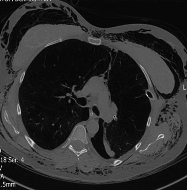
Figure 19.

Severe pneumothorax and surgical emphysema 2 days after insertion of EBVs in the left upper lobe of a 42-year-old lady. The valves are visible in the left hilum successfully occluding the left upper lobe. The lower aspect of a collapsed left upper lobe is also seen. Pneumothorax in this patient did not respond to the intercostal chest drain due to the formation of bronchopleural fistula. Valves needed to be removed. EBV: endobronchial valve.
