
Figure 5. Nivolumab shares similar glycan patterns to mouse aPD-1 and is transferred to macrophages via FcγRs.
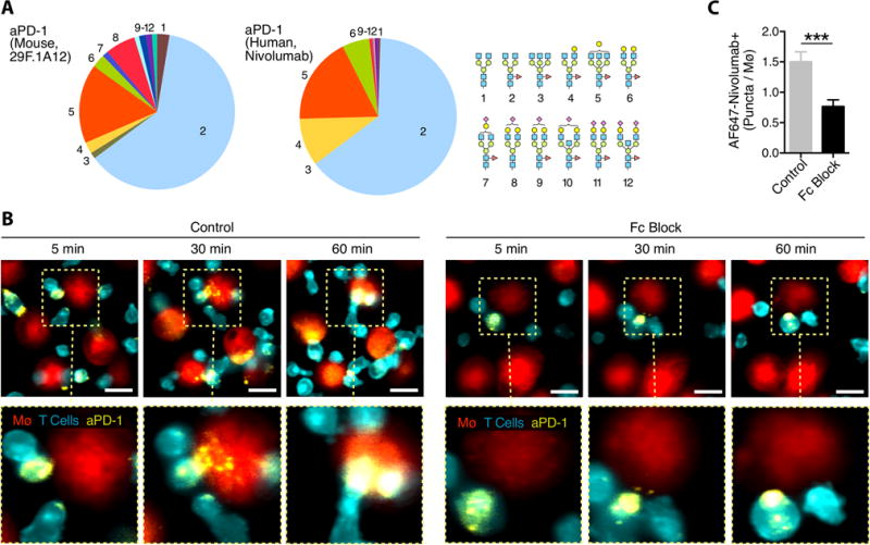
(A) HPLC analysis of the glycan patterns found on the mouse aPD-1 mAb and nivolumab shows the G0F isoform to be predominant, but glycosylation is not uniform. (B) AF647-labeled nivolumab (yellow) was used to stain the surface of aCD3 stimulated PKH-green labeled human CD8+ T cells (cerulean) co-incubated with PKH-red labeled peripheral blood mononuclear cell-derived macrophages in the presence or absence of Fc Block. Scale bars represent 20 μm. (C) Quantification of AF647+ puncta per macrophage confirms that nivolumab is transferred via FcγRs. Values represent SEM for 4 separate experiments.