
Figure 1.
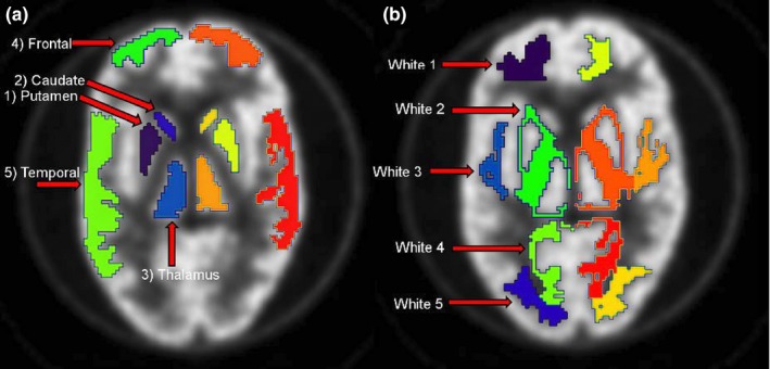
Illustration of VOIs in gray matter for both brain hemispheres in (A) and VOIs in white matter in (B). These VOIs were used to assess RC for different brain structures and regions as function of experimental condition. [Color figure can be viewed at wileyonlinelibrary.com]