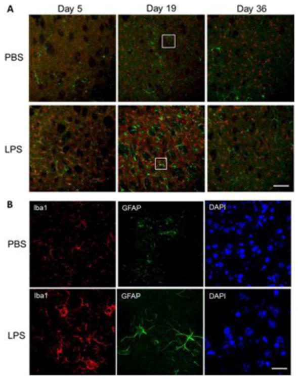
Figure 4.

Activated microglia and astrocytes in the striatum of mice treated with LPS. (A) Immunofluorescence staining for microglia marker ionized calcium-binding adapter molecule 1 (Iba1, red), astrocyte marker glial fibrillary acidic protein (GFAP, green), and DAPI (blue) in striatum on experimental days 5, 19 and 36 after treatment with PBS or LPS. Microglial Iba1 levels were highly increased on day 5 and continued to day 36. (B) Higher magnification at 19 days demonstrates the difference in microglia phenotype and activation in LPS-treated mice. Bar: 500 μm (A) and 50 μm (B).
