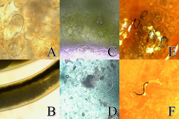
Fig 3. Images of multiple stages of Angiostrongylus cantonensis in situ.
a. Embryonated A. cantonensis eggs in the lung tissue of a rat (40X). b. A. cantonensis eggs that were located in the uterus of a female worm (40X). c. A. cantonensis L1 larvae in lung visualized by tissue squash (10X). d. A. cantonensis L1 larvae observed in rat feces (10x). e. Small black worms, later determined to be L3 A. cantonensis larvae observed in the rat’s lungs. f. The transition between the esophagus and intestine are clearly defined in A. cantonensis L3 larvae.

