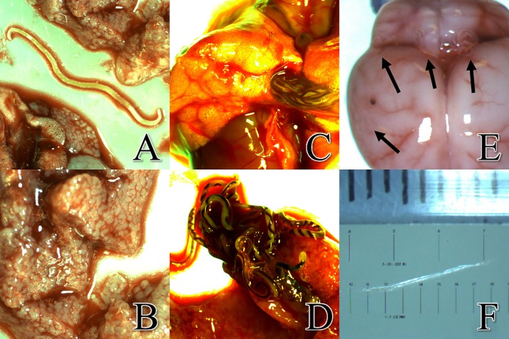
Fig 4. Evidence of infection included presence of decaying encysted adults, granular lung lobes, live adult worms in the pulmonary artery and detection of worms in the brain.
a. Decaying, encysted adult Angiostrongylus cantonensis worms, apparently indicative of a previous infection, were observed in the rat’s lungs. b. Granular upper right lobe of lung. c. Adult A. cantonensis visible in the intact pulmonary artery. d. Adult A. cantonensis emerging from the pulmonary artery of a rat. Males are smaller and females are larger with helical-striped appearance. (See S1 Video). e. Superior view of Rattus exulans brain at a magnification of 6.6X. During dissection, A. cantonensis were commonly found at locations indicated by arrows. The dark spot seen on the surface of the right hemisphere is believed to be a hemorrhage. Hemorrhages were often times an indicator of worm location during dissections. f. Measurement of A. cantonensis L5 from the brain measuring approximately 10 mm. The exsiccated nature of the worm is due to light intensity of the microscope.

