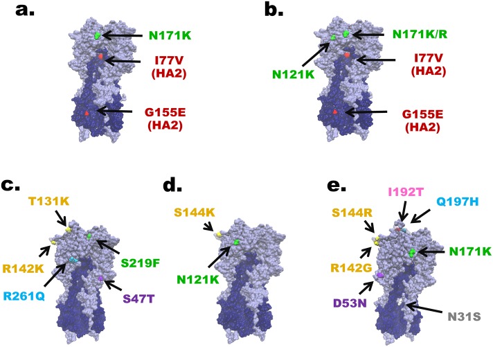
Fig 3. The defining residue substitutions placed on the HA protein structure of A(H3N2).
Differences between A(H3N2) strains in this study and A/Hong Kong/4801/2014 were visualized on the homotrimeric HA structure of A/Aichi/2/1968 (Protein Data Bank accession number: 1HGE). (a) to (e) correspond to groups I to V, respectively. Residues on the five antigenic sites are color-coded: A (yellow), B (pink), C (purple), D (green) and E (blue). Mutation not located within the antigenic site (white) and mutations in HA2 (red) are also noted.

