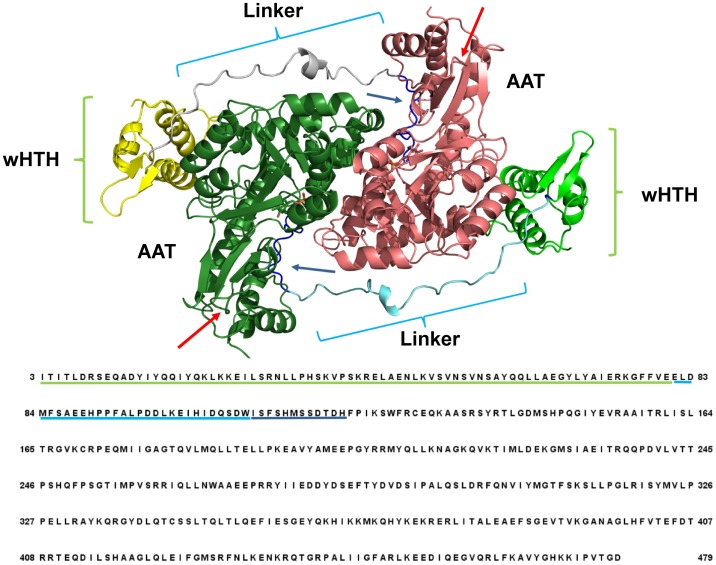
Fig 1. Scheme of the structure of GabR dimer with the corresponding amino acid sequence.
Ribbon colors indicate the different domains in the two subunits: yellow and light green indicate the wHTH domains; cyan and grey the two linkers; dark green and dark pink the AAT domains. Green and cyan underlines map the positions of the wHTH and linker domains onto the sequence, respectively. Dark blue underlines and arrows mark the loop connected to the linker. Cyan and green curly brackets indicate respectively the location of the linkers and HTH domains on the structure. Red arrows mark the position of the two rebuilt loops. Labels ease the visual identification of the domains. Sequence numbering corresponds to that used in the manuscript text.

