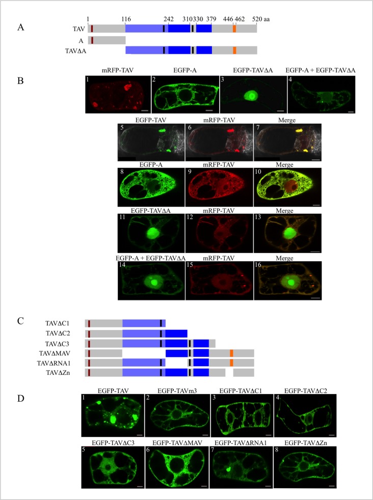
Fig 2. Characterization of CaMV TAV regions involved in the formation of inclusion bodies.
(A, C) Schematic representation of TAV and TAV deletion mutants: A, TAVΔA, TAVΔC1, TAVΔC2, TAVΔC3, TAVΔMAV, TAVΔRNA1 and TAVΔZn. Numbers above the diagram of full-length TAV indicate the amino acids. The functional domains of TAV are depicted as in Fig 1. (B) Transient expression of mRFP-TAV (panel 1), EGFP-A (panel 2), EGFP-TAVΔA (panel 3), and EGFP-A and EGFP-TAVΔA together (panel 4) in tobacco BY-2 cells, and competition assays performed in BY-2 cells co-transfected with plasmids encoding mRFP-TAV and EGFP-TAV (panels 5–7), EGFP-A (panels 8–10), EGFP-TAVΔA (panels 11–13) and both EGFP-A and EGFP-TAVΔA (panels 14–16), respectively. (D) Identification of TAV domains involved in the formation of inclusion bodies by transient expression of EGFP-TAV deletion mutants in BY-2 cells. Observations of TAV and TAV mutants fused to EGFP (B, D) or mRFP (B) were made 16 h after transfection, by LSCM. The LSCM settings and acquisition conditions of the images (single sections) were identical in all (B) and (D) panels. Scale bars: 10 μm.

