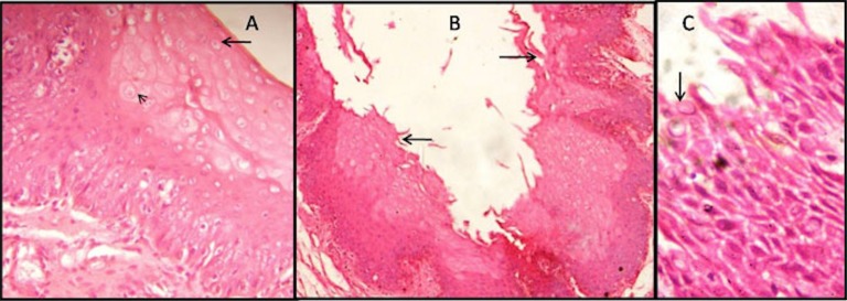
Fig. 2.
Late postmortem changes in the gingiva (8-16hrs, 16-24hrs). (A) Cytoplasimic vacuolation (arrow) and ballooning of cells (arrow head) evident in the superficial and spinous layer of epithelium in 8-16 hrs of PMI (H&E,X400). (B) Shredding (arrow) and other epithelial changes are observed in the whole epithelium in subgroup C (H&E,X100). (C) Disruptive epithelium showing nuclear degenerative changes (arrow) in subgroup C (H&E,X400).

