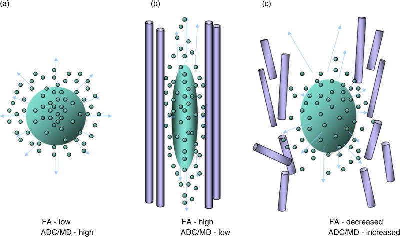
Fig. 1.
(a) This diagram depicts unconstrained molecular movement of a water droplet when there are no membranes to hold the water within. (b) Vertically oriented axon with a membrane wall that constrains the direction of movement of intracellular water as it moves in the same vertical direction (see up-and-down arrows) as the constraining membrane. However, as shown in (c), if the membrane breaks down or is degraded, water molecules disperse in a similar manner to the unconstrained condition. FA = fractional anisotropy, ADC = apparent diffusion coefficient, MD = mean diffusivity. Figure was created by Geri R. Hanten, Ph.D., Baylor College of Medicine.

