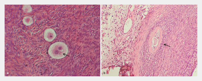
Fig. 4.

Histological sections of the xenograft depicting a multinucleated oocyte as well as an atretic follicle. Extremely rare, multinucleated oocytes were present ( a ; 200 × magnification) inside the xenograft. In addition, some large atretic follicles could be identified ( b ; 100 × magnification).
