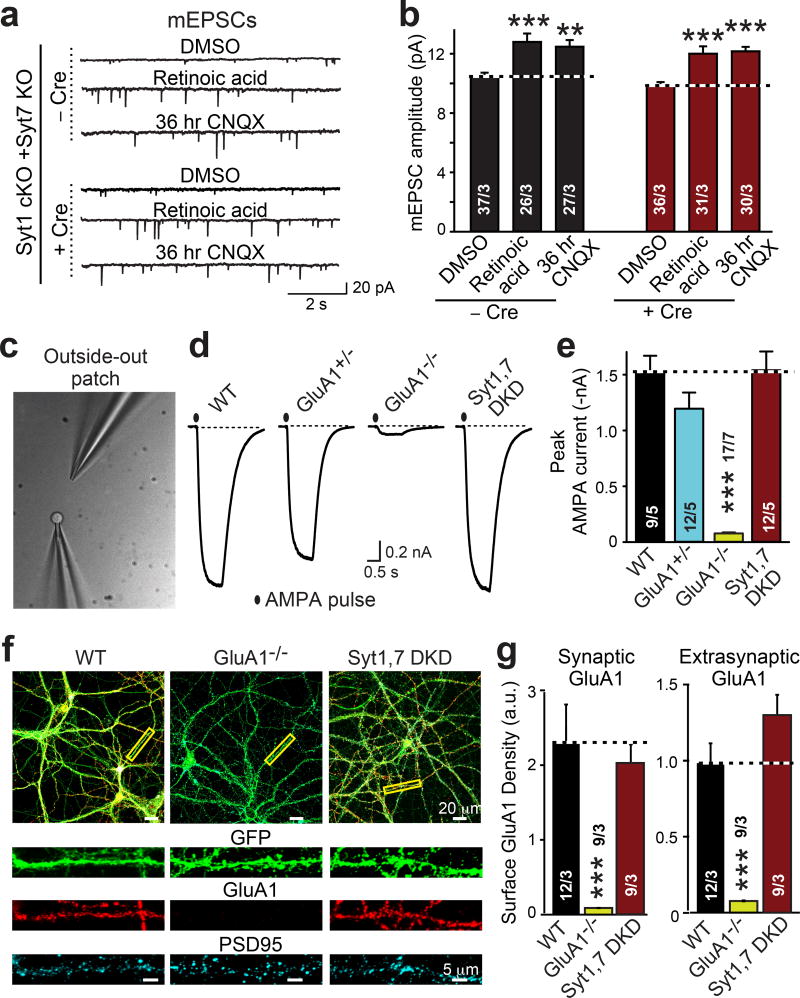
Figure 3. Postsynaptic Syt1,7 deficiency neither decreases AMPAR recruitment during homeostatic plasticity nor surface AMPAR levels.
a & b, Syt1,7 DKO does not impair AMPAR exocytosis induced in cultured hippocampal slices by chronic synaptic silencing with CNQX (36 hr) or by acute application of retinoic acid (4 hr).
c, Phase-contrast image of a patch pipette with a nucleated outside-out patch (left) and an AMPA-puffing pipette (right).
d, Representative traces of currents induced by s-AMPA (10 µM for 1 s) in outside-out patches.
e, Summary graph of the mean AMPA-puff-induced peak current amplitude.
f, Representative images of control (WT), GluA1 KO (GluA1−/−), and Syt1,7 DKD hippocampal neurons that express EGFP (green) and were stained for surface GluA1 receptors (red) and cytoplasmic PSD95 (cyan).
g, Summary graphs of surface GluA1 staining intensity per pixel in synaptic (signal coincident with PSD95) and extrasynaptic membranes (signal non-coincident with PSD95).
Data are means ± SEM (numbers in bars = number of neurons/mice analyzed). Statistical significance was assessed with Kruskal-Wallis ANOVA followed by pairwise comparisons with the Mann-Whitney U test (***, p<0.001).

