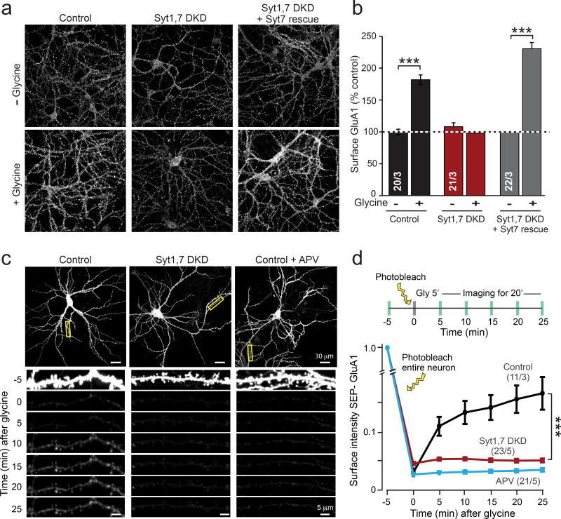
Figure 4. Syt1,7 deficiency blocks AMPAR exocytosis during ‘chemical LTP’ in cultured hippocampal neurons.
a & b, Representative images (a) and summary graphs (b) of GluA1 surface immunostaining as a function of chemical LTP (cLTP) induced by glycine.
c & d, Representative images (c) and summary graph (d) of live-cell SEP-GluA1 fluorescence in hippocampal control neurons expressing transfected SEP-GluA1, imaged before and after cLTP induction with glycine.
Data in b and d are means ± SEM (numbers in bars = number of neurons/independent cultures analyzed). Statistical significance was assessed by Mann-Whitney U test (b) or two-way ANOVA (d; ***p < 0.05).

