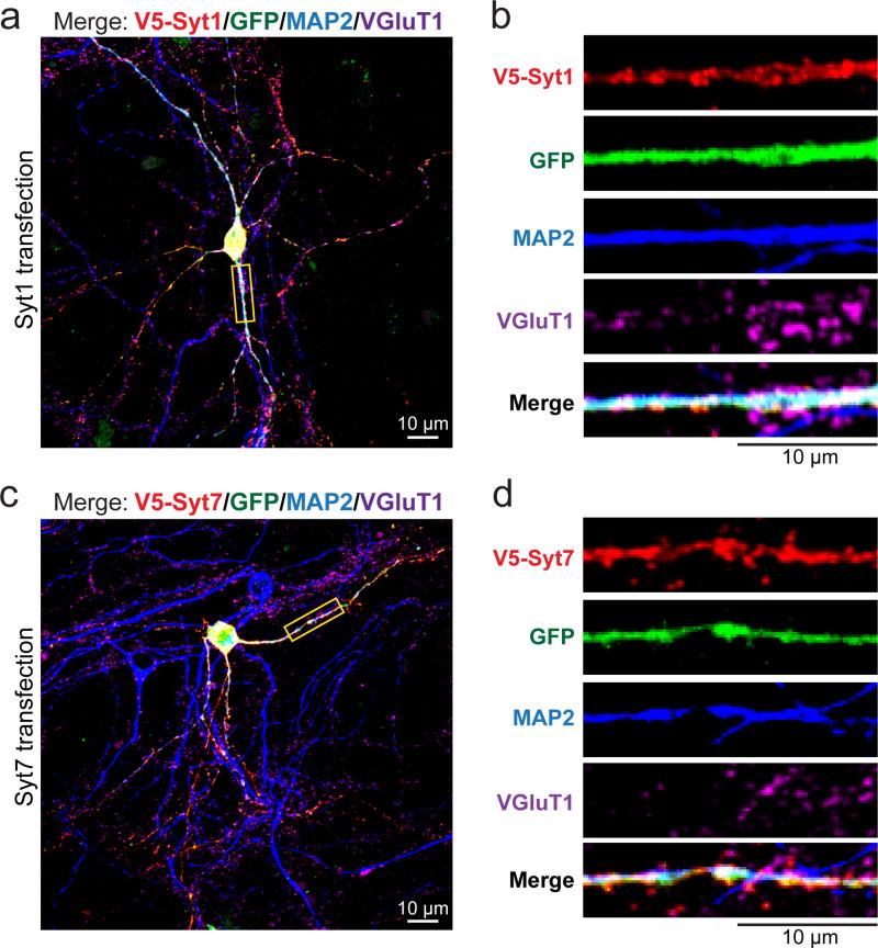
Extended Data Figure 4. Dendroaxonal localizations of Syt1 and Syt7 in cultured hippocampal neurons.
a, Representative image of a WT cultured hippocampal neuron transfected with a vector co-expressing V5-tagged Syt1 and GFP. Neurons were stained for V5 (red), MAP2 (blue), and VGluT1 (magenta) with GFP in green; image shows the merged staining for all four markers.
b, Enlarged images of a segment of the dendrite marked by a yellow box in a, illustrating the distribution of individual markers.
c & d, Same as a & b, but for V5-tagged Syt7.

