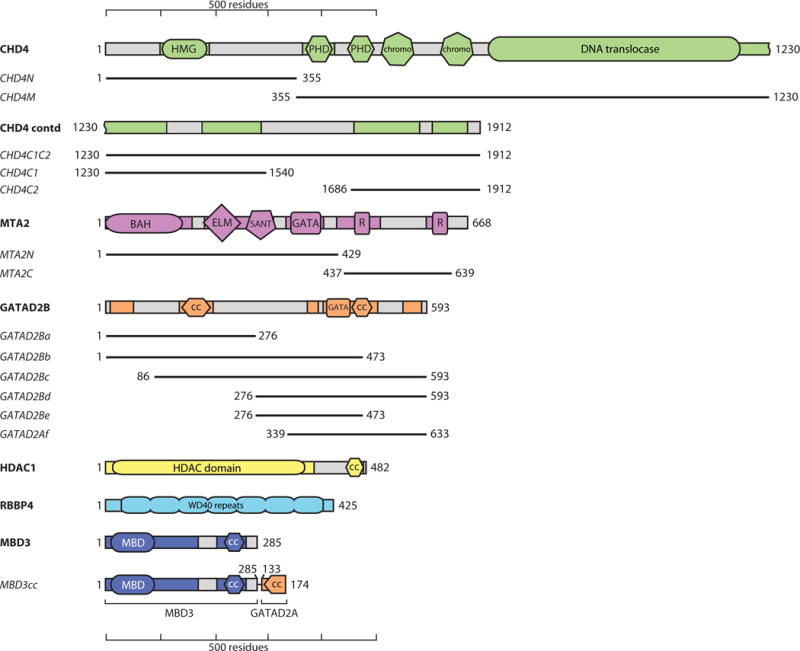
Figure 1. Topology of the NuRD components.

Only one paralogue of each protein is shown. Domains known or predicted to be ordered are shown in colours, and regions predicted to be disordered are shown in grey. Colouring is preserved in other figures. R indicates RBBP-binding motifs. CC indicates coiled coils. Numbering is for the human proteins (UniProt IDs are CHD4: Q14839; MTA2: O94776; GATAD2B: Q8WXI9; HDAC1: Q13547; RBBP4: Q09028; MBD3: O95983). Shorter constructs used in pulldown experiments are indicated with black lines and names in italics. MBD3cc is a construct that fuses MBD3 with the N-terminal coiled-coil domain of GATAD2A. This latter domain forms a dimer with the MBD3 coiled-coil, stabilizing MBD3 [34].
