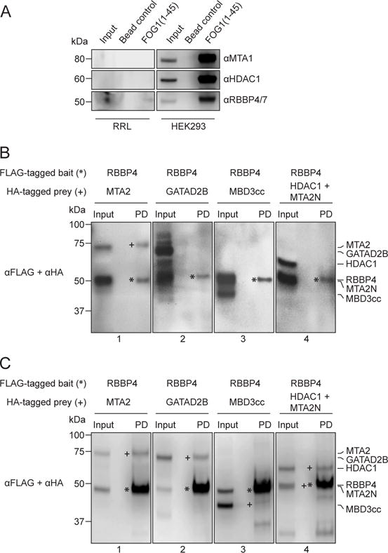
Figure 4. More reliable NuRD subunit interactions can be derived from in vitro rabbit reticulocyte lysate expression.

(a) Western blotting with NuRD-specific antibodies (indicated) shows negligible quantities of several NuRD subunits in rabbit reticulocyte lysates (RRL). As a positive control, all tested NuRD subunits could be readily detected in HEK293 cells. (b) Western blots of pulldowns of proteins co-expressed in RRL, using FLAG-RBBP4 as bait and various HA-tagged prey NuRD subunits as prey. The pulldowns show that RBBP4 interacts with MTA2 (Panel 1) but not GATAD2B (Panel 2), MBD3cc (Panel 3) or HDAC1+MTA2N (Panel 4). Bait proteins have been denoted with *, while the prey proteins, if observed in the pulldown lane (PD), are denoted with +. (c) The same experiment as (b), but with proteins co-expressed in HEK293 cells. In this case, RBBP4 appeared to interact with MTA2 (Panel 1), GATAD2B (Panel 2), MBD3cc (Panel 3) and HDAC1+MTA2N (Panel 4).
