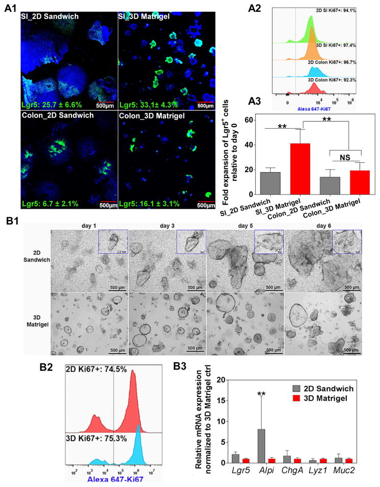
Figure 6.
(A) BLT Sandwich culture is capable of expanding freshly isolated mouse SI, colon crypts and (B) patient-derived SI crypts. Equal number of freshly isolated crypts were evenly seeded into 2D Sandwich and 3D Matrigel systems and cultured for 4 days (mouse) or 6 days (human). A1: Confocal images showing the morphology and Lgr5-GFP expression of SI and colonic IECs in 2D and 3D systems. Percentage of Lgr5-GFP+ cells was noted inside each picture in green. A2: Flow cytometry comparing Ki67 expression of IECs expanded from both systems. A3: Comparison of the fold expansion of mouse SI and colonic Lgr5-GFP+ cells between 2D and 3D systems. Mean ± SEM, n = 4 donor mice each with 3–5 wells/group. B1: Photographs showing the proliferation and morphological change of human SI IECs cultured under both systems for 6 days. Insets show the growth of a representative single crypt into a monolayer patch with diameter ~ 1 mm at day 6 (scale bar: 100 μm). B2: Flow cytometry comparing Ki67 expression between both systems at day 6 (representative from n = 3 patients). B3: qPCR analysis comparing the mRNA expression of key IEC lineage markers between both systems at day 6. Mean ± SEM, n = 3 trials using freshly isolated SI crypts from three patients. ** p<0.01: significantly different between 2D and 3D cultures.

