Figure 3.

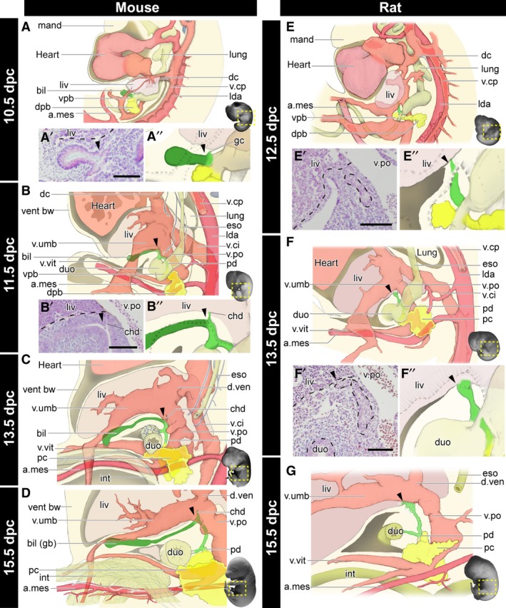
Morphogenesis of the biliary system in the mouse and rat. All panels are left lateral views. See also Figs 4 and S2 for the histological sections, digitally reconstructed models and latex‐injected models. (A–D) Developmental scheme of the hepatobiliary system in mice. The biliary bud (green), pancreas (yellow), artery (magenta), and vein (pink) are drawn. The gallbladder–cystic duct (Gb–Cd) domain (dark green) can be identified as the distal domain from the branching point (arrowheads) of the common hepatic duct (chd). Details of the common hepatic ducts are shown in panels (A’), (A’’) and (B’), (B’’). (E–G) The developmental scheme in rats. The arrowheads indicate the landmarks comparable with mice. The Gb–Cd domain is clearly absent. a.mes, arteria mesenterica cranialis; bil, biliary bud; chd, common hepatic duct; dc, ductus cuvieri; dpb, dorsal pancreatic bud; duo, duodenum; d.ven, ductus venosus; eso, oesophagus; gb, gallbladder; gc, glandular cord; int, intestine; lda, left dorsal aorta; liv, liver or liver primordium; lung, lung bud; mand, mandibular process; pc, pancreas; pd, pancreatic duct; v.ci, vena cava inferior; v.cp, vena cava posterior; vent bw, ventral bodywall; vpb, ventral pancreatic bud; v.po, vena portae; v.umb, vena umbilcailis; v.vit, vena vitelline. Scale bar: 500 μm. [Correction added on 24 October 2017, after first online publication: the abbreviations cited on this figure was added on figure caption]
