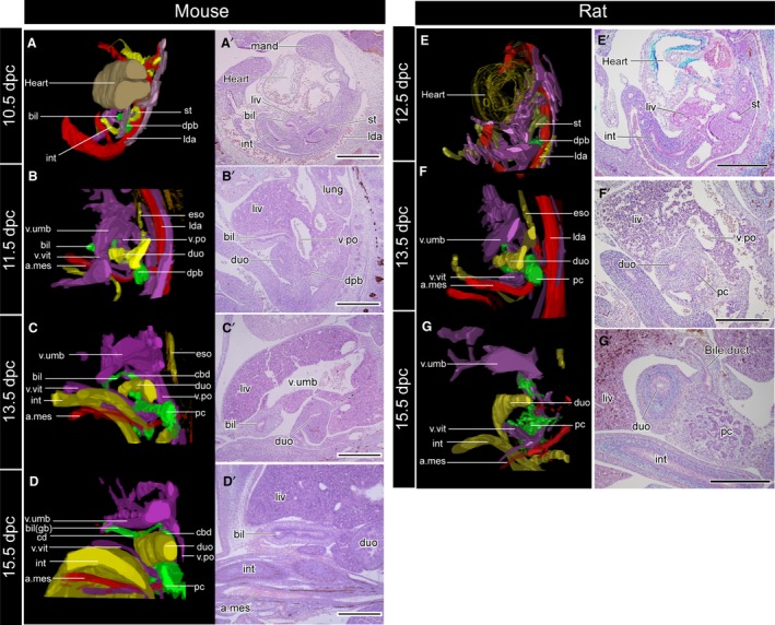
Figure 4.

Three‐dimensional reconstructed models (A–D) and the underlying histological sections (A′–D′). The topographical relationships of all samples are the same as in Fig. 3. The sections were subjected to immunohistochemical (IHC) staining for acetylated tubulin to visualise the peripheral nerves. The biliary tract is innervated by the intestinal part of the vagus, but the nerve supplies had not been formed at the developmental stages examined in the present study. a.mes, arteria mesenterica cranialis; bil, biliary bud; cbd, common bile duct; cd, cystic duct; dpb, dorsal pancreatic bud; duo, duodenum; eso, oesophagus; gb, gallbladder; int, intestine; lda, left dorsal aorta; liv, liver or liver primordium; lung, lung bud; mand, mandibular process; pc, pancreas; v.po, vena portae; v.umb, vena umbilcailis; v.vit, vena vitelline. Scale bar: 500 μm. [Correction added on 24 October 2017, after first online publication: the abbreviations cited on this figure was added on figure caption]
