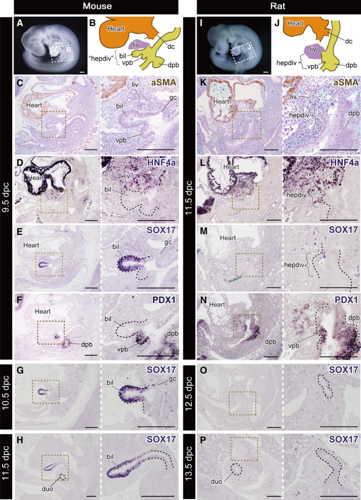
Figure 5.

Molecular patterning of the hepatobiliary primordia. All panels are left lateral views. (A) The mouse embryo at 9.5 dpc. The scheme of the hepatobiliary primordia is shown in (B). ‘hepdiv’ in (B) indicates the presumptive domain that corresponds to the hepatic diverticulum of the 11.5 dpc rat in (J). (C–F) Histological sections of the same 9.5‐dpc embryo with immunohistochemical (IHC) staining for aSMA, HNF4a, SOX17 and PDX1. (G,H) Sections of 10.5‐ and 11.5‐dpc mice stained for SOX17. The SOX17‐positive domain is localised in the distal portion of the biliary bud. (I) A rat embryo at 11.5 dpc. The scheme of the hepatobiliary primordia is shown in (J). (K–N) Sections of the same 11.5‐dpc embryo with IHC staining in the same manner as the mouse. (M,N) Sections of 12.5‐ and 13.5‐dpc rats stained for SOX17. The black dotted line in the higher‐magnification panels indicates the extrahepatic biliary tract. bil, biliary bud; dc, ductus cuvieri; dpb, dorsal pancreatic bud; duo, duodenum; gc, glandular cord; hepdiv, hepatic diverticulum; liv, liver or liver primordium; vpb, ventral pancreatic bud. Scale bars: 200 μm. [Correction added on 24 October 2017, after first online publication: the abbreviations cited on this figure was added on figure caption].
