Figure 5.

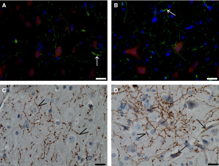
TH immunostaining in the claustrum. (A, B) Immunofluorescent endothelial cells (white arrows) and axons (green) contacting cell bodies (NeuN, red). (C, D) Immunoperoxidase reaction showing TH‐ir puncta and axons with varicosities running at all directions. Arrowheads indicate terminals that surrounded and defined cell bodies. Black arrows indicate positive endothelial cells. Scale bars = 10 μm.
