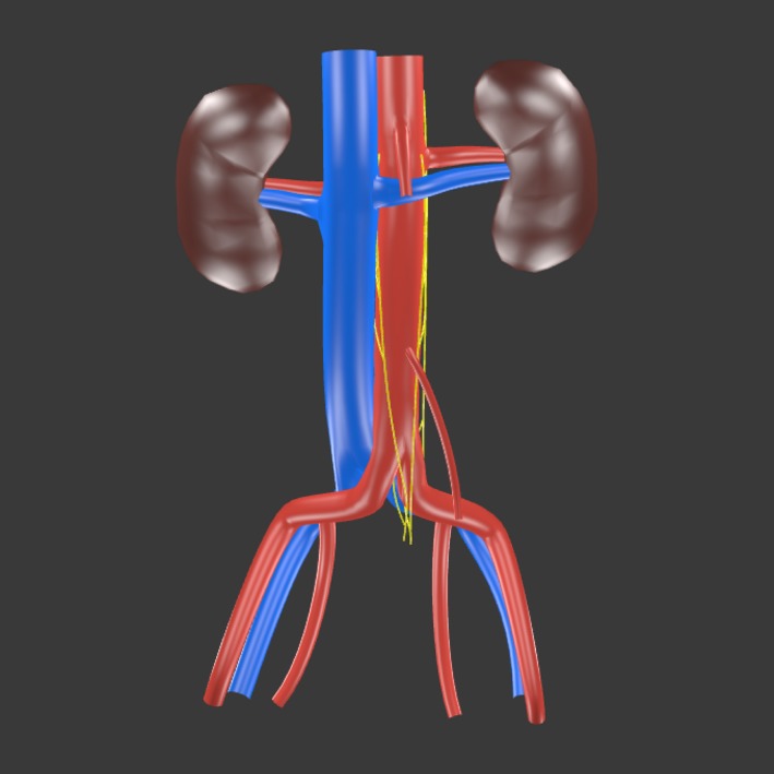
Figure 3.

A 3D model (click to activate) showing the average position and course of the infrarenal lumbar splanchnic nerves (LSNs) as determined from the present study. The rostrocaudal positions of the LSNs are drawn to scale relative to the infrarenal length. Adobe Acrobat Reader is required to interact with the 3D model.
