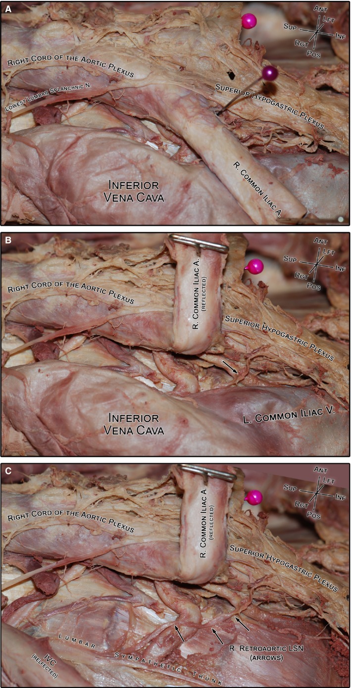
Figure 5.

A photograph of the gross dissection of a typical right retroaortic lumbar splanchnic nerve (LSN; specimen #1854). (A) The course of the retroaortic LSN cannot be clearly observed without moving the vasculature. For reference, the purple pin indicates the bifurcation of the aorta. (B) Reflecting the right common iliac artery reveals the underlying retroaortic nerve (black arrow) and its connection with the superior hypogastric plexus. (C) Lateral translation of the inferior vena cava reveals the entire course of the retroaortic nerve from the lumbar sympathetic trunk to the superior hypogastric plexus. IVC, inferior vena cava.
