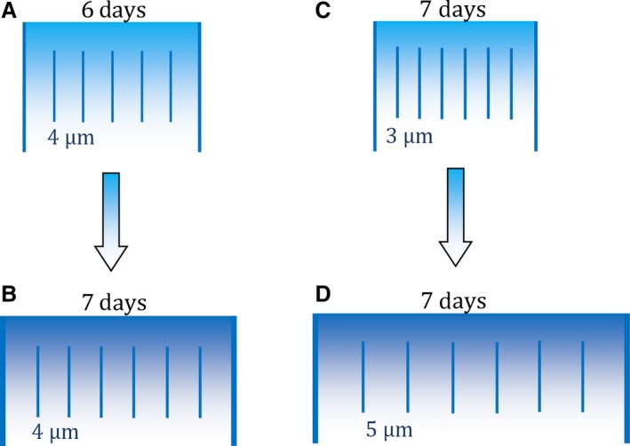
Figure 5.

Cell mechanisms underlying different thicknesses of human enamel layers. Long lines illustrate two adjacent Retzius lines (enamel layer) in one outer lateral region of the crown. Short lines represent daily cross‐striations with either 3, 4 or 5 μm of enamel between adjacent striations, representing different daily enamel secretion rates (DSR). Retzius periodicity (RP) increases from 6 days in (A) to 7 days in (B). Layer (B) increases in width because ameloblasts have secreted enamel for an extra day, relative to (A), but the DSR remains constant (e.g. this study; Mahoney et al. 2017). Another developmental mechanism is illustrated by (C) and (D). RP remains the same in both illustrations. Layer (D) increases in width because of the greater DSR, relative to (C), which might be expected when deciduous incisors are compared with second molars along the tooth row of the same individual (Mahoney, 2015).
