Fig. 5.
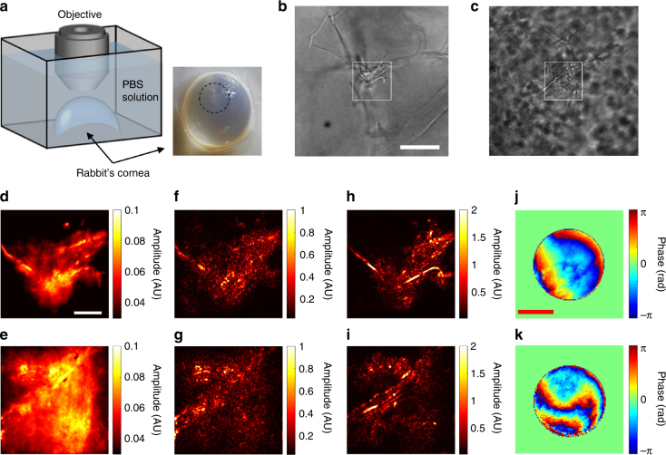
Experimental demonstration of CLASS microscopy for imaging the hyphae of Aspergillus cells in a rabbit’s cornea. a Layout of the sample geometry. A rabbit’s cornea infected by A. fumigatus was immersed in PBS solution in a Petri dish and placed under the objective lens. The black dashed circle in the photo indicates the infection site. b, c Transmission images taken by illuminating the cornea from the bottom of the Petri dish in (a) using a light emitting diode (λ = 780 nm). Scale bar, 40 µm. d, e Incoherent addition of time-gated and angle-dependent reflection images for the white rectangular boxes are shown in (b, c) respectively. Scale bar, 10 µm. f, g Images taken by CASS microscopy in the epi-detection geometry for the white rectangular boxes are shown in (b, c) respectively. h, i Same as (f, g) respectively, but after applying CLASS algorithm. Color bar, intensity in arbitrary unit. j, k Specimen-induced aberration maps identified during the acquisition of (h, i) respectively. Scale bar, k 0 α. Color bar, phase in radians

