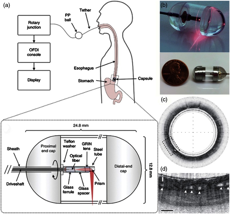
Fig. 17.
Tethered capsule OCT endomicroscopy. (a) Overview of the device. The tether allows retrieval of the capsule, which also contains the drive shaft and optical fiber, after being swallowed through the esophagus and passing briefly into the stomach of the subject. Inset: schematic of the capsule. (b) Close-up of capsule with scanning mechanism active and positioned adjacent to a US penny for scale. (c) In vivo tethered capsule endomicroscopy image obtained from a patient with histopathologically confirmed Barrett’s esophagus. (d) Threefold () expanded view showing an irregular luminal surface, heterogeneous backscattering, and glands within the mucosa (arrowheads). Tick marks in (c) represent 1 mm. Scale bars represent 0.5 mm. Reprinted with permission from Macmillan Publishers Ltd.123

