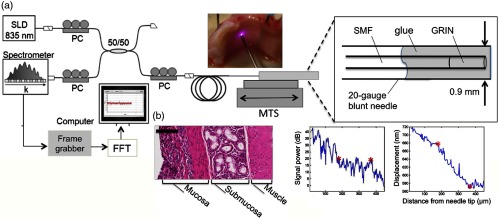
Fig. 20.
Tissue boundary detection using OCT-elastography in a handheld needle probe. (a) Schematic of forward facing needle probe and OCT system. Abbreviations: SLD, superluminescent diode; PC, polarization controller; MTS, motorized translation stage; SMF, single-mode fiber; GRIN, gradient-index fiber. (b) Boundary detection in ex vivo porcine tracheal wall with comparative histology. Scale bar represents . Reprinted with permission.138

