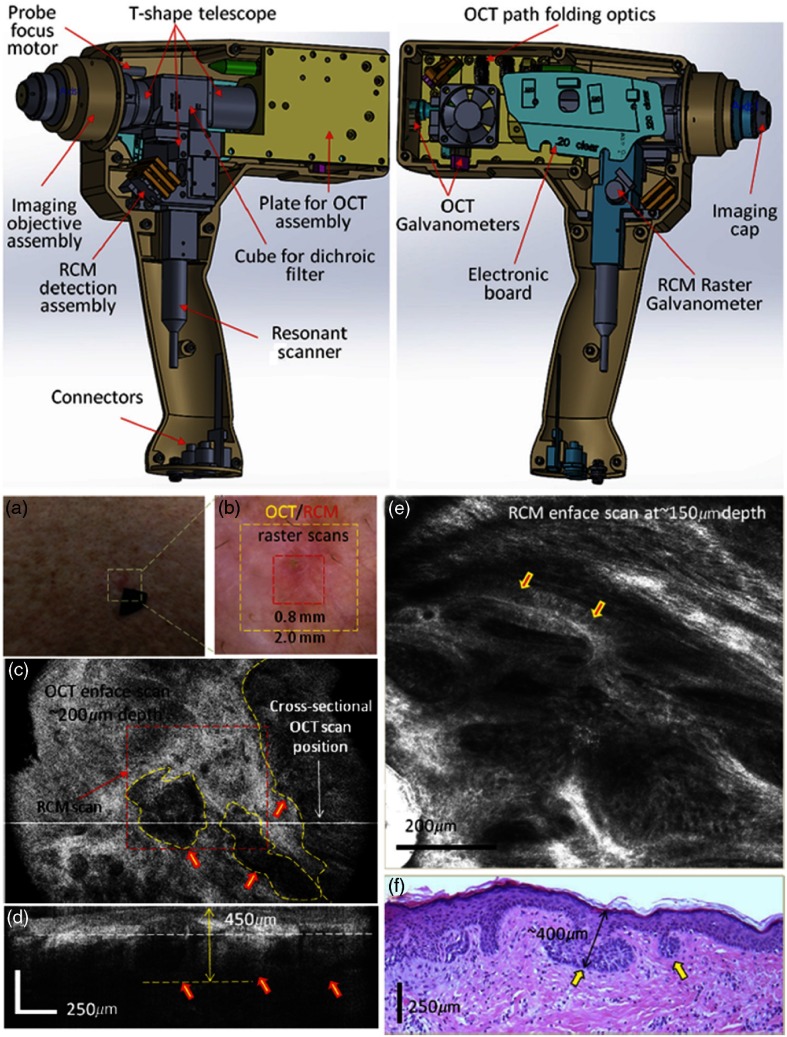
Fig. 26.
Handheld OCT-RCM probe. (Top) CAD design and schematic. (Bottom) (a) Clinical image of an erythematous macule. (b) Dermoscopy image of shiny white lines and serpentine vessels in the imaging region. (c) En face and (d) cross-sectional OCT images of hypoechoic areas (arrows), suggestive of BCC. (e) RCM showing cord-like structures with peripheral palisading (arrows) admixed with a fibrotic stroma, suggestive of BCC. (f) Histology of the lesion confirms superficial BCC, with multiple small tumor nests originating from the epidermis (H&E, ). Modified and reprinted with permission.183

