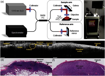
Fig. 3.
Second-generation intraoperative portable spectral-domain OCT clinical system. (a) OCT system schematic and (b) photo of the OCT system. Physical dimensions were greatly reduced over the previous version. (c, e) Representative intraoperative OCT and (d, f) corresponding histopathology images of a normal, nonmetastatic (left) and cancerous metastatic (right) ex vivo human LNs. All scale bars represent 0.5 mm. Modified and reprinted with permission.53

