Figure 1.
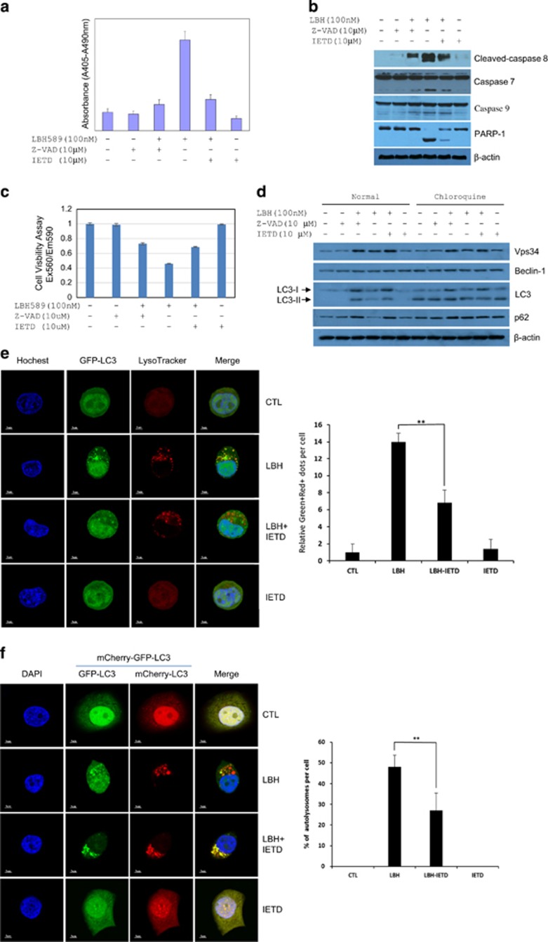
Inhibition of caspase 8 promotes the fusion of autophagosome with lysosome. (a) Inhibition of LBH589-induced apoptosis by caspase inhibitors. Apoptotic cell death of MCF-7 cells induced by treatment with the indicated concentrations of LBH589 and/or caspase inhibitors for 48 h. For caspase inhibitor treatment, cells were pretreated with 10 μM z-VAD-FMK (Z-VAD) or z-IETD-FMK (IETD) for 4 h. (b) Z-VAD or IETD inhibited LBH589-induced cleavage of caspase 7, caspase 8, caspase 9 and PARP-1 in MCF-7 cells treated as indicated for 24 h. (c) Cell viability of MCF-7 cells treated as indicated for 24 h. (d) Accumulation of Vps34, LC3 and p62 in MCF-7 cells treated as indicated with or without 20 μM chloroquine for 24 h. (e) Confocal microscopic evaluation of autophagosome-lysosome fusion in MCF-7 cells. MCF-7 cells stably expressing EGFP-LC3 (Green) treated as indicated for 18 h were stained with lysosome tracker staining (Red). Immunofluorescence analyses were performed using confocal microscopic detection (63 × oil). Left, representative confocal images. Right, relative Green+red+ dots per cell calculated from 20 cells; bars, s.d. **P<0.01. (f) Disruption of autophagosome-lysosome fusion in MCF-7 cells. MCF-7 cells stably expressing tfLC3 were treated as indicated for 18 h. Immunofluorescence analyses were performed using confocal microscopic detection (60 × oil). Left, representative confocal images. Right, % of autolysosomes per cell was calculated from 20 cells by using the formula of (1-yellow dots/red dots) × 100; bars, s.d. **P<0.01. DAPI, 4', 6-diamidino-2-phenylindole.

