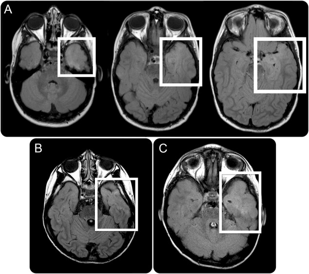
Figure 1. MRI of patient 1 with fluid-attenuated inversion recovery signal.
(A) Axial MRI of patient 1 demonstrating diffuse cortical/subcortical high fluid-attenuated inversion recovery (FLAIR) signal confined to the anterior left superior temporal gyrus and mesial temporal structures (white boxes). (B–C) Subtle increased FLAIR signal in the anterior temporal lobes of patients 2 and 3, respectively, suggestive of possible cortical dysplasia.

