Figure 5.
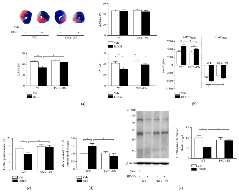
PKCε contributed to HDGF-induced reduction of reperfusion injury. PKCε dominant negative mice (PKCε-DN) and wild-type (WT) littermates with or without 50 μg/kg recombinant mouse HDGF treatment intramyocardially were subjected to 45 min of cardiac ischemia followed by 24 h reperfusion. (a) Ratio of area at risk to left ventricle area (AAR/LV), ratio infarct size to AAR ratio (I/AAR), and ratio of infarct size to LV area (I/LV) of hearts were assessed. Data represent mean ± SEM of values from five mice. (b) The maximum rates of rise and decline of left-ventricular pressure (+dp/dtmax and −dp/dtmax) assessed at 24 h reperfusion. Data are mean ± SEM of values from six mice. (c) Quantitative analysis of TUNEL positive cells at 24 h reperfusion. Data are mean ± SEM of values from three hearts per group, with at least 3000 nuclei examined per heart. (d) Mitochondria were isolated from heart tissue after reperfusion injury and the activity of ALDH2 in mitochondria was measured. Data represent mean ± SEM of values from three mice. (e) 4-HNE protein adducts in heart tissues was assessed. Data represent mean ± SEM of values from three mice. ∗P < 0.05.

