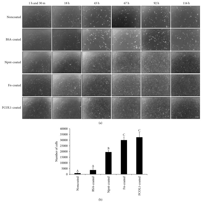
Figure 2.
Microscopic observation and cell number counting. (a) hDPSCs were seeded into PBS-, BSA-, Npnt-, Fn-, or FCOL1-coated 24-well plates (non-PS) at the concentration of 4 × 10³/well in DMEM supplemented with 10% FBS, penicillin/streptomycin (50 U/mL; 50 μg/mL) (scale bar: 200 μm). Cell morphology was observed at 1 h and 30 m, 18 h, 43 h, 67 h, 92 h, and 116 h. (b) hDPSCs were seeded into 24-well plates (non-PS) at the concentration of 4 × 10³/well in the same culture media as illustrated in (a), cell number was counted at day five. Different symbols represent significant differences, p < 0.01 by post hoc Tukey's HSD test.

