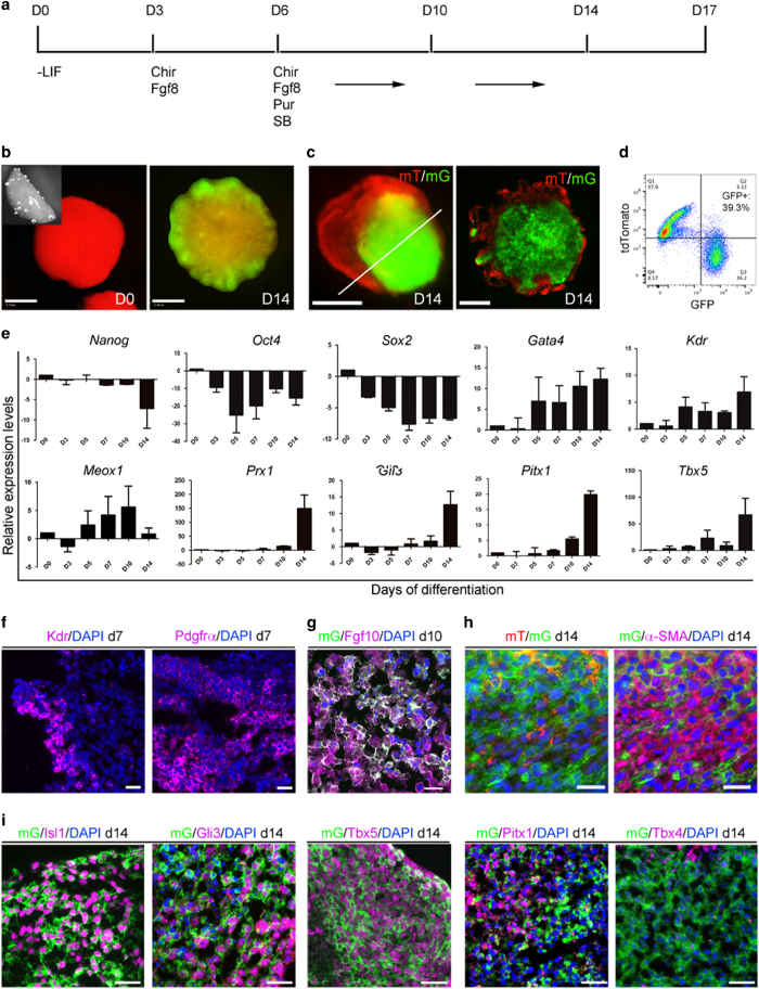
Figure 3.
Derivation of limb progenitor-like cells from 3D FB culture of Prx1Cre:mT/mG iPSCs. (a) Diagram of the protocol used in this study. LIF (leukemia inhibitory factor), Chir (CHIR99021, 3 μM), Fgf8 (10 ng/ml), Pur (Purmorphamine, 4 μM), SB (SB431542, 2 μM). (b) Examples of FBs freshly made and cultured for 14 days (d14). Inset in d0 is a side view of the hemispherical FB immediately after transfer to culture medium. Multiple budding structures are GFP+ in a d14 FB (b). (c) A smaller bud-like structure, with a GFP+ core covered by tdTomato+ outside layer, revealed in cross-section. (d) Flow cytometry analysis of GFP+ cells in D14 FBs, showing about 40% cells were induced to switch on GFP expression. (e) Real-time PCR detection of gene expression in FBs, for pluripotency markers (Nanog, Oct4, Sox2), lateral plate mesoderm markers (Gata4, Kdr, Meox1) and limb field related genes (Prx1, Gli3, Pitx1, Tbx5). Gene expression levels were normalized to glyceraldehyde 3-phosphate dehydrogenase (GAPDH), and compared with d0 specimens. Results were from three independent experiments. (f) Detection of mesoderm markers Kdr and Pdgfrα at d7 of differentiation. (g) Detection of Fgf10 in day 10 FB cultures. (h) Induced GFP+ cells expressed mesodermal marker α-SMA (in purple), detected by immunofluorescence. (i) Detection of Isl1, Gli3, Tbx5, Pitx1 and Tbx4 in induced GFP+ cells by immunofluorescence. Scale bars: (b): 0.5 mm, (c): 200 μm, (f–i): 20 μm.

