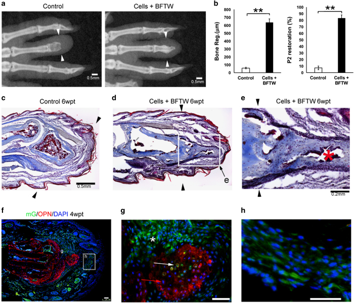
Figure 5.
Stimulated adult mouse D3P2 regeneration after transplantation of iPSC-derived limb progenitor-like cells. (a) Examples of adult mouse D3P2 with or without limb progenitor-like cell +BFTW transplantation), by X-ray imaging. (b) Statistical analysis of length of bone regeneration and P2 restoration in adult D3P2 with graft (limb progenitor-like cells+BFTW) or without (control). Error bars: standard error, N=9. **P<0.01, Student’s t-test. (c–e) Sections of D3P2 with Trichrome staining, in control (d) or cells+BFTW group (d, e), 6 wpt. Black arrows indicate amputation levels. White dotted boxes indicate areas shown in (e). (e) An enlarged view of the P2 regenerate, showing well-integrated outgrowth of the P2 bone, and marrow formed in the regenerated bone (red asterisk). (f, g) Contribution of GFP+ iPSC-derived limb progenitor cells (exemplified by the white arrow) and GFP− host cells (exemplified by the red arrows) in the bone regenerate (marked by OPN expression, in red). *Indicates areas of GFP+ cells outside the bone. (h) Contribution of GFP+ iPSC-derived limb progenitor cells in the connective tissues of the adult D3P2. Scale bars: 0.5 mm in (a, c, d), 0.2 mm in (e), 50 μm in (g, h).

