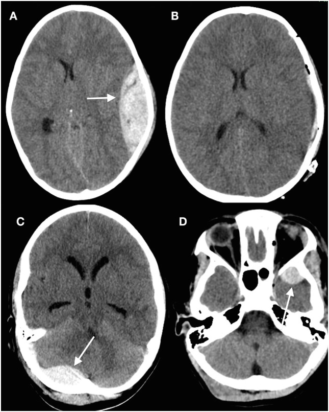
Figure 2.

Epidural hematomas occur in a variety of locations. (A) Head computed tomography (CT) showing a typical convexity epidural hematoma in a child; (B) evacuated hematoma in the same patient; (C) posterior fossa epidural hematoma (arrowed) underlying a suboccipital fracture; (D) epidural hematoma anterior to the left temporal tip (arrowed) (13) (modified).
