
Fig. 2.
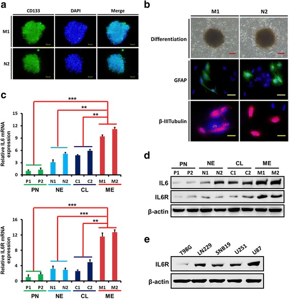
The expression of IL6 and IL6R in patient-derived primary glioma cells. a–b Neurospheres comprised of CD133-positive glioma stem-like cells (GSCs) obtained from primary cultures (a) became adherent and differentiated into GFAP- or β III tubulin -positive cells (b). Scale bars: red = 50 μm, yellow = 25 μm. c-d qPCR (c) and western blotting analyses (d) showed that, compared with the classical, neural, and proneural glioma cells, the levels of IL6 and IL6R mRNA and protein expression were higher in mesenchymal glioma cells. e The expression of IL6R in different glioma cell lines was examined by western blotting. **P < 0.01, ***P < 0.001. Classical, CL; neural, NE; proneural, PN; mesenchymal, ME