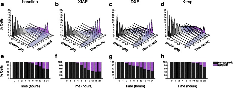
Fig. 4.

Distribution of cPARP concentration in population-level model. a-d: Histogram showing the percentage of the 2000 cells with a given cPARP concentration, in response to 10 nM TSP1 stimulation. (a) Baseline model; (b) XIAP downregulation; (c) DXR treatment; and (d) Increased nuclear translocation rate. A different color is assigned to each time point. The cPARP threshold is marked by a solid line and the region in the x-y plane beyond the threshold is shaded as light purple. (e-h): The predicted percentage of non-apoptotic (black) and apoptotic (purple) cells in response to 10 nM TSP1 stimulation. (e) Baseline model; (f) XIAP downregulation; (g) DXR treatment; and (h) Increased nuclear translocation rate
