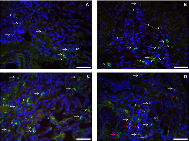
FIGURE 2.

Immunofluorescence staining for tryptase, chymase, and DAPI nuclear staining (40×) forali the four groups (A: early normal allograft, B: late normal allograft, C: acute cellular rejection, D: chronic lung allograft dysfunction) for patient #3. DAPI nuclear staining shows as blue color indicating any nucleated cell. Tryptase only positive cells (MCT cells) show green-colored granules (white arrows) which appear to increase from figure A to D. MCTC cells show dual staining (red and green, red arrows) which appear to be significantly increased in (D) representing the CLAD group.
