
Figure 4. Comparative analysis of receptor binding to Makona and Mayinga GPs.
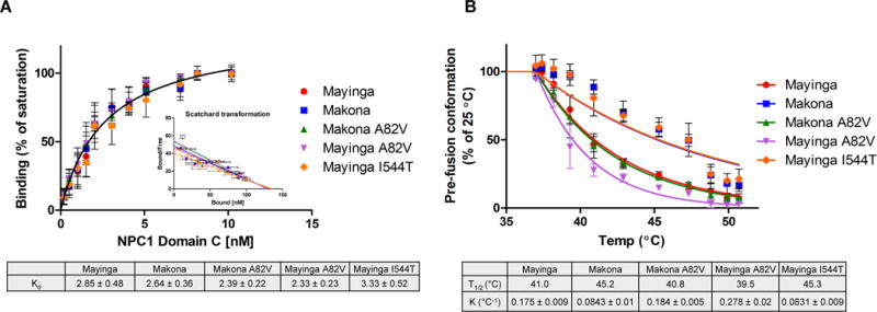
A. MLV particles pseudotyped with the indicated GPs were cleaved to expose the NPC1 binding domain and bound to plates using KZ52 mAb. Bound particles were incubated with increasing concentrations of purified NPC1 Domain C-His6. Bound NPC1 Domain C was detected using α-His6 antibody. Binding is reported as mean ± s.d. (n = 4) of the % of maximum binding for each GP. Pairwise comparison indicates the affinities of these GPs for NPC1 Domain C are not statistically significant (p = 0.18). See also Figure S3.
B. MLV particles pseudotyped with the indicated GPs were cleaved and then heated for 30min at the specified temperatures (T = 25–51.7°C). After cooling to room temperature, virus particles were captured on ELISA plates coated with the GP2 prefusion conformation-specific mAb KZ52. Results are mean ± s.d. (n = 3) of the % of binding for each GP at T = 25°C. Makona vs Makona A82V: p = <0.0001; Mayinga vs Mayinga A82V: p = <0.0001; Mayinga vs Mayinga I544T: p = <0.0001; Mayinga vs Makona A82V: p = 0.4; Makona vs Mayinga I544T: p = 1. See also Figure S5.