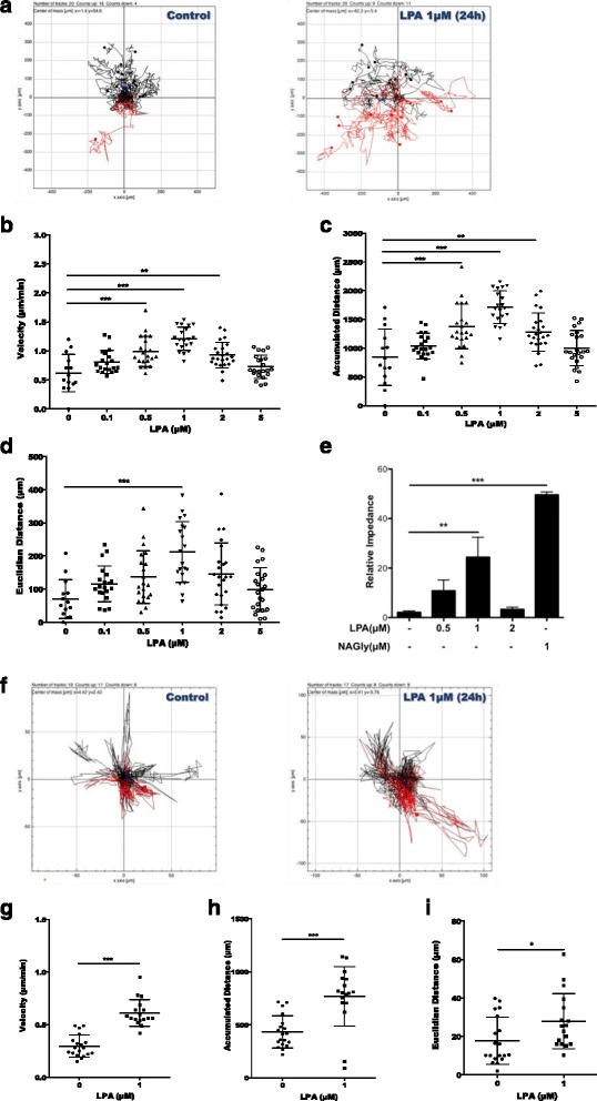
Fig. 10.

LPA induces microglial chemokinesis and chemotaxis. a BV-2 cells were cultured in 24-well plates, serum-starved overnight, and incubated with 0.1% BSA or with different LPA concentrations for 24 h. Migration was analyzed by time-lapse microscopy. b Velocity, c accumulated distance, and d Euclidean distance of at least 20 cells per sample were determined by ImageJ. e Chemotaxis was analyzed using the xCELLigence system. Serum-starved cells were allowed to migrate across uncoated Transwell inserts (CIM plates) for 24 h. Chemotaxis [0.1% BSA or LPA (1 μM) added to the lower compartment] was followed in real time by continuous electrical impedance measurement. NAGly (1 μM) was used as positive migration control. f PMM were cultured on PDL-coated 24-well plates, serum-starved overnight, and treated with 0.1% BSA or LPA (1 μM). Time-lapse microscopy was used to analyze 2D migration of at least 20 viable cells per sample per condition. g Velocity, h accumulated distance, and i Euclidean distance were determined using ImageJ. For BV-2 cells, results from three independent experiments performed in triplicate were expressed as mean + SD (**p < 0.01, ***p < 0.001; one-way ANOVA with the Bonferroni correction LPA-treated versus untreated). For PMM, the results from two experiments in triplicate are shown as mean + SEM (*p < 0.05, ***p < 0.001; unpaired Student’s t test)
