
Fig. 3.
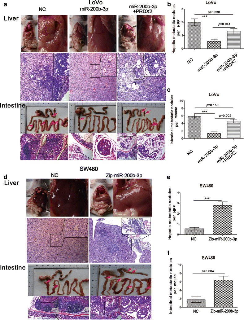
MiR-200b-3p inhibits CRC cells’ metastatic capacity by targeting PRDX2 in vivo. a Intestinal and hepatic metastatic nodules after subcutaneous tumors derived from LoVo/NC, LoVo/miR and LoVo/miR + PRDX2 cells were transplanted in the mesentery at the distal end of cecum in mice (n = 5) for 6 weeks. Red arrows point at potential metastatic nodules in intestines. Scale bars represent 50 μm. b, c The number of hepatic metastatic nodules (b) or intestinal metastatic nodules (c) of mice with tumors derived from LoVo/NC, LoVo/miR and LoVo/miR + PRDX2 cells. The number of hepatic metastatic nodules per mouse was counted under the microscope, with five high power fields (HPF) observation (***p < 0.001). d Intestinal and hepatic metastatic nodules after subcutaneous tumors derived from SW480/NC and SW480/Zip-miR cells were transplanted in the mesentery at the distal end of cecum in mice (n = 5) for 6 weeks. Red arrows point at potential metastatic nodules in intestines. Scale bars represent 50 μm. e, f The number of hepatic metastatic nodules (e) or intestinal metastatic nodules (f) of mice with tumors derived from SW480/NC and SW480/Zip-miR cells. The number of hepatic metastatic nodules per mouse was counted under the microscope, with five HPF observation (***p < 0.001)