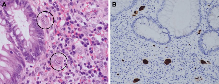
Figure 1.
Pathological presentations of CMV colitis.
Notes: CMV colitis was diagnosed by both CMV inclusion bodies and positive IHC staining in the colonic tissue. (A) H&E stain (40× objective) showed typical intranuclear (owl’s eye) and intracytoplasmic (eosinophilic punctiform) CMV inclusions within the circles. (B) IHC stain (20× objective) was performed with 1:200 diluted Novocastra™ lyophilized mouse monoclonal antibody against CMV pp65 antigen and showed strong focal CMV immunoreactivity with brownish areas.
Abbreviations: CMV, cytomegalovirus; H&E, hematoxylin and eosin; IHC, immunohistochemistry.

